
ファイル:Cytokinesis-electron-micrograph.jpg
Cytokinesis-electron-micrograph.jpg (745 × 451 ピクセル、ファイルサイズ: 200キロバイト、MIME タイプ: image/jpeg)
ウィキメディア・コモンズのファイルページにある説明を、以下に表示します。
|
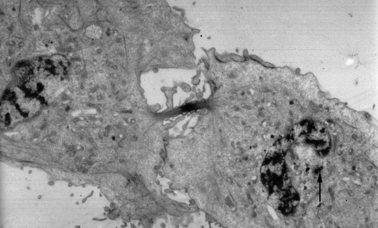
Picture from English Wikipedia
An electron micrograph image of a cell that has almost completed cell division and cytokinesis. Mitosis has already been completed. An arrow points to a centrosome still present near one of the nuclei.
From http://www.wadsworth.org/bms/SCBlinks/web_mit2/RES_MIT.htg/teleoph.jpg アーカイブされたコピー at the Wayback Machine, the Wadsworth Center, which is part of the New York State Department of Health and devoted to public education. Since it's part of the US government, I'll assume public domain.
このファイルは、アメリカ合衆国の連邦政府と雇用関係にある公務員がその職務上作成したアメリカ合衆国政府の著作物であり、アメリカ合衆国の著作権法上パブリックドメインに属します (17 U.S.C. §105)。
注意:このライセンスは、アメリカ合衆国政府の著作物についてのみ効力を有します。アメリカ合衆国の各州、郡、その他の地方自治体が作成した著作物に対しては適用できません。
|
 | |
| このファイルは著作権法の既知の制約(隣接権や関連する権利を含む)から自由であると特定されています。 | ||
Uploaded 07:21, 21 July 2005 .by user . Natalinasmpf . . 745x451 (87580 bytes) (An electron micrograph image of a cell that has almost completed cell division and cytokinesis. Mitosis has already been completed. An arrow points to a centrosome still present near one of the nuclei.
このファイルは、アメリカ合衆国の連邦政府と雇用関係にある公務員がその職務上作成したアメリカ合衆国政府の著作物であり、アメリカ合衆国の著作権法上パブリックドメインに属します (17 U.S.C. §105)。
注意:このライセンスは、アメリカ合衆国政府の著作物についてのみ効力を有します。アメリカ合衆国の各州、郡、その他の地方自治体が作成した著作物に対しては適用できません。
|
| |
| このファイルは著作権法の既知の制約(隣接権や関連する権利を含む)から自由であると特定されています。 | ||
)
ファイルの履歴
過去の版のファイルを表示するには、その版の日時をクリックしてください。
| 日付と時刻 | サムネイル | 寸法 | 利用者 | コメント | |
|---|---|---|---|---|---|
| 現在の版 | 2011年5月24日 (火) 12:54 | | 745 × 451 (200キロバイト) | Zephyris | Reverted to version as of 12:52, 24 May 2011 |
| 2011年5月24日 (火) 12:54 |  | 745 × 451 (200キロバイト) | Zephyris | Reverted to version as of 12:50, 24 May 2011 Reversion seemed not to work | |
| 2011年5月24日 (火) 12:52 |  | 745 × 451 (200キロバイト) | Zephyris | Reverted to version as of 12:50, 24 May 2011 Confusion with cached images | |
| 2011年5月24日 (火) 12:52 |  | 745 × 451 (200キロバイト) | Zephyris | Oops, uploaded the original file last time by accident! | |
| 2011年5月24日 (火) 12:50 |  | 745 × 451 (200キロバイト) | Zephyris | Inverted image: It is more common to show more intensly absorbing features in an electron micrograph (e.g. chromatin and the midbody) as dark rather than light. Asjusted levels and contrast: To both use the full histogram range and emphasise detail in the | |
| 2005年12月1日 (木) 17:43 |  | 745 × 451 (86キロバイト) | Rasbak | Picture from English Wikipedia An electron micrograph image of a cell that has almost completed cell division and cytokinesis. Mitosis has already been completed. An arrow points to a centrosome still present near one of the nuclei. |
ファイルの使用状況
以下のページがこのファイルを使用しています:
グローバルなファイル使用状況
以下に挙げる他のウィキがこの画像を使っています:
- ar.wikipedia.org での使用状況
- bn.wikipedia.org での使用状況
- ca.wikipedia.org での使用状況
- en.wikipedia.org での使用状況
- es.wikipedia.org での使用状況
- gl.wikipedia.org での使用状況
- ht.wikipedia.org での使用状況
- hy.wikipedia.org での使用状況
- it.wikipedia.org での使用状況
- kk.wikipedia.org での使用状況
- nl.wikipedia.org での使用状況
- nl.wikibooks.org での使用状況
- pl.wikipedia.org での使用状況
- pt.wikipedia.org での使用状況
- ru.wikipedia.org での使用状況
- sh.wikipedia.org での使用状況
- sl.wikipedia.org での使用状況
- sr.wikipedia.org での使用状況
- tr.wikipedia.org での使用状況
- uk.wikipedia.org での使用状況